EEG
EEG stands for electroencephalogram. An EEG measures the brain's electrical activity using electrodes placed on the head. These electrodes are small metal plates with wires attached. The wires can be glued to the scalp, or sometimes a special cap with built-in electrodes is used. Additionally, a few electrodes are often placed on other parts of the body, such as the shoulder. In most cases, video recording is used during the test to assist in reviewing the results.
Different Types of EEG Examinations
There are various types of EEG tests, differing in duration and location. An EEG examination can take place in different settings, such as the Clinical Neurophysiology (KNF) department, the Epilepsy Monitoring Unit, the inpatient ward, or even at home. In some cases, patients need to keep a seizure diary, while in others, it is not required. The doctor determines which type of EEG examination is necessary for you.
24-Hour EEG Monitoring
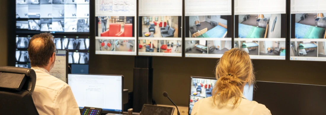
A commonly used EEG test involves a one-day hospital stay. The 24-hour EEG is designed to answer various epilepsy-related questions, such as confirming a diagnosis or evaluating the effectiveness of a medication treatment. The goal of this test is to observe precisely what happens in the brain during recording and to correlate this with external symptoms. In many cases, it is not necessary to trigger an actual seizure, though specific methods to provoke seizures may be used based on individual circumstances.
During a 24-hour EEG monitoring, you will:
Stay with a few fellow patients in a monitoring area (a lounge-like space) or a private suite.
Have continuous brain activity monitoring via EEG.
Be observed by lab technicians and nurses with video cameras.
Continue EEG monitoring overnight in your own bedroom or suite.
Direct Brain Recording
Direct brain recording is considered when epilepsy surgery might be a treatment option, but there is still uncertainty about the precise location of the seizure focus.
This advanced pre-surgical seizure monitoring helps doctors pinpoint with greater accuracy the exact area of the brain responsible for seizures. Additionally, it helps define the boundaries of regions that control essential bodily functions. There are three types of direct brain recording:
Strip Electrodes – Electrodes on thin wires are placed directly on the brain's surface in the suspected seizure area under general anesthesia.
Grid Electrodes – Electrodes on a small mat (a few square centimeters in size) are placed directly on the brain in the suspected seizure area. Seizure activity is monitored in the hospital for a period of time. If epilepsy surgery is deemed feasible, the surgery follows the removal of the electrode mat.
Stereo-EEG (sEEG) – Electrodes on thin wires are inserted into the brain via a needle under general anesthesia. This seizure monitoring can last several weeks and takes place at Kempenhaeghe. If the results confirm that epilepsy surgery is a treatment option, the operation is scheduled a few months after the removal of the wires.
Before undergoing this extensive examination, you will receive detailed personal information from a neurologist and/or specialized nurse regarding the procedure and practical considerations.
More Information
Before your EEG test, you will be provided with detailed information about the type of EEG you will undergo, how the examination will proceed, and any practical aspects to consider.
Laboratory and blood tests
For most patients at Kempenhaeghe, blood tests are focused on monitoring medication levels. Sometimes, blood tests are needed to determine the cause of your symptoms.
The concentration of medication in the blood provides insight into how well the medication is working. Each person requires a dosage tailored to them. The combination with other medications can also influence blood levels. For most patients, a low blood concentration means that the medication is not effective enough, while a high blood concentration increases the risk of side effects. Blood tests can also be conducted to check for possible medication side effects (such as liver function, electrolyte levels, etc.).
Your doctor can tell you more about the purpose of the blood test. In Heeze, blood tests are conducted at Kempenhaeghe’s in-house (prick) laboratory. In Breda, blood samples must be taken at a different location.
Would you like more information about Kempenhaeghe’s laboratory services, for example, to learn what happens to blood and tissue samples after testing?
MRI
MRI Examination
MRI scans allow doctors to determine whether an abnormality in the brain could be the cause of your symptoms.
MRI stands for Magnetic Resonance Imaging. It produces highly detailed images of the brain using magnetic fields. An MRI scan generates a series of cross-sectional images of the head and brain. This allows the doctor to identify potential abnormalities such as damage, scarring, developmental disorders, or tumors.
What Happens During an MRI?
An MRI examination is painless and requires no special preparation. During the MRI at Kempenhaeghe, you will lie on an examination table with a plastic holder placed over your head. It is important to remain still. A laboratory technician can communicate with you via a microphone inside the MRI tunnel.
During the scan, you will hear a tapping sound. You will be given earplugs or headphones with music to reduce the noise. When the noise stops, one image sequence has been completed. Capturing a single sequence takes anywhere from 30 seconds to over 10 minutes, and most MRI examinations consist of multiple sequences.
Before the scan, you will receive detailed information about the specific type of MRI you will undergo, how the examination will proceed, and any practical considerations.
Functional MRI (fMRI)
MRI scans allow doctors to determine whether an abnormality in the brain could be the cause of your symptoms. A functional MRI (fMRI) can show which areas of the brain are active during specific tasks or at rest.
While lying inside the MRI scanner, you will perform small tasks. This provides the doctor with information about the brain regions responsible for movement and cognitive functions such as memory, language, and planning.
For a small percentage of patients, fMRI is combined with an EEG examination, known as an EEG-fMRI. Before undergoing this test, you will receive personalized information about the procedure.
Epilepsy Sugery Check
Epilepsy Surgery Check
Are you a mentally competent adult patient with difficult-to-treat epilepsy, where at least two types of anti-seizure medication have failed to provide sufficient control? In that case, epilepsy surgery may be a solution. Successful epilepsy surgery can significantly improve your quality of life. To quickly assess—within four weeks—whether epilepsy surgery has a good chance of success for you, we offer the Epilepsy Surgery Check.
One-Day Intake and Examinations
Your treating neurologist can refer you to our expertise center for the Epilepsy Surgery Check. The process starts at our Heeze location with an intake consultation with a neurologist specialized in epilepsy. On the same day, you will undergo an MRI scan and a 24-hour EEG. The results of your MRI scan are compared with our extensive MRI database, a process known as post-processing.
Results Consultation
Within four weeks, you will have a results consultation at Kempenhaeghe. Our neurologist will then share the findings with your own neurologist. Together with your neurologist, you will discuss whether epilepsy surgery is a suitable option for you. You will remain under the care of your own hospital throughout the process.
Neurological Examination
Neurological Examination
(Pediatric) neurological examination consists of an extensive conversation with the (pediatric) neurologist about the reason for the visit and, if necessary, a physical examination to check the main functions of the nervous system. The neurological examination is not burdensome. The (pediatric) neurologist observes and tests the senses, motor skills, and coordination and assesses reflexes.
Neuropsychological and Psychological Examination
A neurological disorder (such as epilepsy) may be accompanied by problems with language, memory, or concentration. The condition may also impact a person's self-image or mood. It is not uncommon for questions to arise regarding the acceptance of the consequences of the condition in areas such as living, working, parenting, learning, and relationships.
This may lead to a referral to a psychologist at Kempenhaeghe. Various psychologists with specific areas of expertise work at Kempenhaeghe. For both children and adults, (neuro-)psychological examination helps identify the concerns. Based on the findings, recommendations are made, for example, regarding further support.
Psychological examinations consist of an intake (in the case of children, also with the parents) and the administration of test research. The tests include answering questions, solving tasks, measuring reaction speed, and conducting memory and attention tests. Additionally, aspects such as mood, personality, and acceptance of the neurological disorder may be examined. The tests are conducted over one or two sessions by a psychological assistant. In some cases, the psychologist is present for part of the time. With your consent, video recordings may be made to support the diagnostic process.
The psychologist evaluates the test results and discusses the findings with you. After the examinations and the results discussion, you will receive a copy of the examination report.
Before the (neuro)psychological examination, you will receive information about the course of the examination, practical considerations, and information about rights and responsibilities related to the examination.